missing translation for 'onlineSavingsMsg'
Learn More
Learn More
Invitrogen™ CD19 Monoclonal Antibody (HIB19), NovaFluor™ UV 765, eBioscience™
Mouse Monoclonal Antibody
€ 176.00 - € 388.00
Specifications
| Antigen | CD19 |
|---|---|
| Clone | HIB19 |
| Concentration | 0.6 μg/Test |
| Content And Storage | 4°C, store in dark, DO NOT FREEZE! |
| Applications | Flow Cytometry |
| Product Code | Brand | Quantity | Price | Quantity & Availability | |||||
|---|---|---|---|---|---|---|---|---|---|
| Product Code | Brand | Quantity | Price | Quantity & Availability | |||||
|
30245068
|
Invitrogen™
H004T02U01-A |
25 Tests |
€ 176.00
Pack of 25 |
Please sign in to purchase this item. Need a web account? Register with us today! | |||||
|
30245179
|
Invitrogen™
H004T03U01-A |
100 Tests |
€ 388.00
Pack of 100 |
Please sign in to purchase this item. Need a web account? Register with us today! | |||||
Description
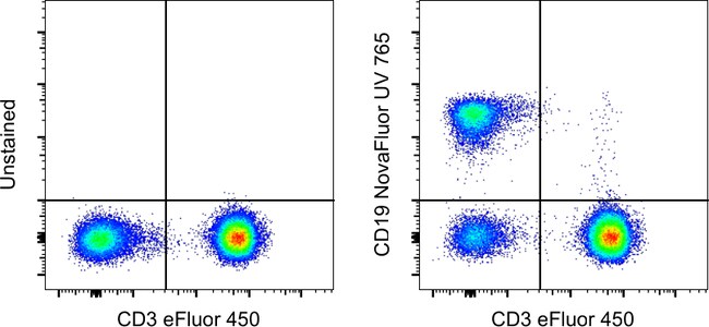
Description: The HIB19 monoclonal antibody reacts with human CD19, a 95 kDa transmembrane glycoprotein. The SJ25C1 antibody and the HIB19 monoclonal antibody recognize overlapping epitopes. This product contains 1 vial of NovaFluor conjugate and 1 vial of CellBlox Plus Blocking Buffer. Applications Tested: This HIB19 antibody has been pre-titrated and tested by flow cytometric analysis of normal human peripheral blood cells. This can be used at 5 μL (0.6 μg) per test. A test is defined as the amount (μg) of antibody that will stain a cell sample in a final volume of 100 μL. Cell number should be determined empirically but can range from 10^5 to 10^8 cells/test. Master mixes. Whole Blood compatibility: When utilizing whole blood (as opposed to density-gradient-purified PBMC), we recommend lysing red blood cells in bulk prior to staining with NovaFluor conjugates. Staining of whole blood with NovaFluor conjugates followed by lysis of red blood cells may result in higher-than-expected background staining.
CD19 is a member of the immunoglobulin superfamily, characterized by two Ig-like domains, and is expressed on B cells throughout all stages of development, excluding terminally differentiated plasma cells. It is also expressed on follicular dendritic cells and has been observed on myeloid leukemia cells, particularly those of monocytic lineage. CD19 is considered the earliest and broadest B cell-restricted antigen, and its expression is found in all B cell precursor leukemias. CD19 forms a multimolecular complex with CD21, CD81, Leu13, MHC class II, and the B cell receptor (BCR), playing a crucial role in B cell signaling. As a signal-amplifying coreceptor for the BCR, CD19 lowers the threshold for antigen receptor-dependent stimulation, allowing B cells to respond specifically and sensitively to various antigens through low-affinity antigen receptors. Signaling through CD19 induces tyrosine phosphorylation, calcium flux, and proliferation of B cells. Beyond its role as a BCR coreceptor, CD19 can also signal independently of BCR co-ligation, serving as a central regulatory component upon which multiple signaling pathways converge. This makes CD19 an important functional regulator of both normal and malignant B cell proliferation. Mutations in the CD19 gene can result in hypogammaglobulinemia, a condition characterized by low levels of immunoglobulins, while CD19 overexpression can lead to B cell hyperactivity. CD19 is expressed on 100% of peripheral B cells, as defined by the expression of kappa or lambda light chains, underscoring its significance in B cell function and immune regulation.Specifications
| CD19 | |
| 0.6 μg/Test | |
| Flow Cytometry | |
| NovaFluor UV 765 | |
| Mouse | |
| RUO | |
| PBS with BSA and 0.09% sodium azide; pH 7.2 | |
| P15391 | |
| 930 | |
| Primary | |
| Affinity chromatography |
| HIB19 | |
| 4°C, store in dark, DO NOT FREEZE! | |
| Monoclonal | |
| Liquid | |
| IgG1 κ | |
| Human | |
| Cd19 | |
| AW495831; B4; B-lymphocyte antigen CD19; B-lymphocyte surface antigen B4; Cd19; CD19 antigen; CD19 molecule; CVID3; differentiation antigen CD19; Leu-12; T-cell surface antigen Leu-12 | |
| Cd19 | |
| Antibody |
Spot an opportunity for improvement?Share a Content Correction
Product Content Correction
Your input is important to us. Please complete this form to provide feedback related to the content on this product.
Product Title