missing translation for 'onlineSavingsMsg'
Learn More
Learn More
Invitrogen™ CD1c Monoclonal Antibody (L161), Super Bright™ 436, eBioscience™

Mouse Monoclonal Antibody
€ 342.00
Specifications
| Antigen | CD1c |
|---|---|
| Clone | L161 |
| Concentration | 5 μL/Test |
| Content And Storage | 4°C, store in dark, DO NOT FREEZE! |
| Applications | Flow Cytometry |
Description
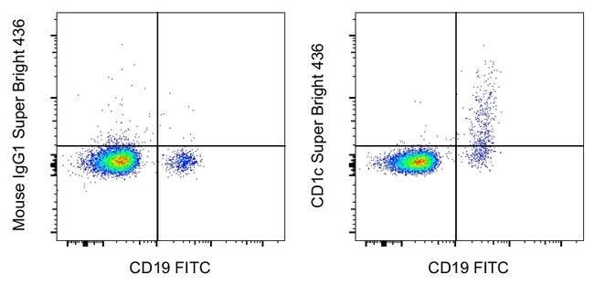
Description: This L161 monoclonal antibody detects CD1c (also known as BDCA-1), a glycoprotein that is noncovalently linked to beta-2 microglobulin on thymocytes and antigen presenting cells such as dendritic and Langerhans cells. This molecule is also expressed on some circulating and marginal zone B cells, as well as in lymph nodes and germinal centers. CD1c is involved in the presentation of lipid antigens such as microbial fatty acids to effector T cells during the adaptive immune response. Finally, alternative splicing gives rise to three different isoforms of CD1c (soluble, membrane, and cytoplasmic/soluble isoforms). Applications Reported: This L161 antibody has been reported for use in flow cytometric analysis. Applications Tested: This L161 antibody has been pre-diluted and tested by flow cytometric analysis of normal human peripheral blood cells. This may be used at 5 μL (0.25 μg) per test. A test is defined as the amount (μg) of antibody that will stain a cell sample in a final volume of 100 μL. Cell number should be determined empirically but can range from 10^5 to 10^8 cells/test. Super Bright 436 can be excited with the violet laser line (405 nm) and emits at 436 nm. We recommend using a 450/50 bandpass filter, or equivalent. Please make sure that your instrument is capable of detecting this fluorochrome. When using two or more Super Bright dye-conjugated antibodies in a staining panel, it is recommended to use Super Bright Complete Staining Buffe...
CD1c is a member of the CD1 family of transmembrane glycoproteins, which are structurally related to major histocompatibility complex (MHC) proteins and form heterodimers with beta-2-microglobulin. This family of proteins is involved in the presentation of lipid and glycolipid antigens, both of self and microbial origin, to T cells during the adaptive immune response. CD1c is expressed on some circulating and marginal zone B cells, as well as in lymph nodes and germinal centers. It plays a crucial role in presenting lipid antigens, such as microbial fatty acids, to effector T cells. The protein encoded by the CD1c gene localizes to late endosomes and lysosomes, utilizing a tyrosine-based motif in its cytoplasmic tail for targeting. Vesicular acidification is required for CD1c to bind lipid antigens effectively. The human genome contains five CD1 family genes organized in a cluster on chromosome 1, with each member differing in cellular localization and specificity for particular lipid ligands. CD1c undergoes alternative splicing, resulting in three different isoforms: soluble, membrane-bound, and cytoplasmic/soluble isoforms, highlighting its functional diversity in immune processes.Specifications
| CD1c | |
| 5 μL/Test | |
| Flow Cytometry | |
| Super Bright 436 | |
| Mouse | |
| RUO | |
| PBS with BSA and 0.09% sodium azide; pH 7.2 | |
| P29017 | |
| 911 | |
| Primary | |
| Affinity chromatography |
| L161 | |
| 4°C, store in dark, DO NOT FREEZE! | |
| Monoclonal | |
| Liquid | |
| IgG1 κ | |
| Human | |
| CD1C | |
| BDCA1; canCD1c; CD1; CD1A; CD1C; CD1C antigen, c polypeptide; CD1c molecule; cortical thymocyte antigen CD1C; differentiation antigen CD1-alpha-3; R7; RP11-101J8.3; T-cell surface glycoprotein CD1c | |
| CD1C | |
| Antibody |
Spot an opportunity for improvement?Share a Content Correction
Product Content Correction
Your input is important to us. Please complete this form to provide feedback related to the content on this product.
Product Title