missing translation for 'onlineSavingsMsg'
Learn More
Learn More
Invitrogen™ CD223 (LAG-3) Monoclonal Antibody (3DS223H), Brilliant Ultra Violet™ 563, eBioscience™, Invitrogen™

Mouse Monoclonal Antibody
€ 185.00 - € 375.00
Specifications
| Antigen | CD223 (LAG-3) |
|---|---|
| Clone | 3DS223H |
| Concentration | 5 μL/Test |
| Applications | Flow Cytometry |
| Classification | Monoclonal |
| Product Code | Brand | Quantity | Price | Quantity & Availability | |||||
|---|---|---|---|---|---|---|---|---|---|
| Product Code | Brand | Quantity | Price | Quantity & Availability | |||||
|
17847040
|
Invitrogen™
365-2239-41 |
25 Tests |
€ 185.00
Pack of 25 |
Please sign in to purchase this item. Need a web account? Register with us today! | |||||
|
17857040
|
Invitrogen™
365-2239-42 |
100 Tests |
€ 375.00
Pack of 100 |
Please sign in to purchase this item. Need a web account? Register with us today! | |||||
Description
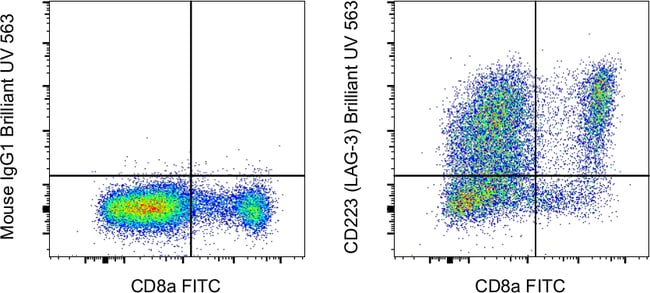
This 3DS223H mAb recognizes human CD223 also known as Lymphocyte Activation Gene 3 (LAG-3). LAG-3 is a 70-kDa surface glycoprotein belonging to the Ig superfamily with homology to CD4. LAG-3 binds to MHC class II with higher affinity than CD4 and is thought to be involved in the negative regulation of T cell activation and homeostatic proliferation. Surface expression of LAG-3 has been reported on activated T cells (including regulatory T cells) and NK cells. CD8+ T cells usually express LAG-3 at significantly higher levels than CD4+ T cells. Coexpression of LAG-3 and CD49b has been proposed to identify human and mouse Type 1 regulatory T cells (Tr1 cells). This 3DS223H antibody will recognize a formaldehyde-fixed epitope. Applications Tested: This 3DS223H antibody has been pre-diluted and tested by flow cytometric analysis of stimulated normal human peripheral blood cells. BUV563 is a tandem dye that emits at 564nm and is intended for use on cytometers equipped with an Ultraviolet (355nm) laser. Please make sure that your instrument is capable of detecting this fluorochrome. When using two or more Super Bright, BV, BUV, or other polymer dye-conjugated antibodies in a staining panel, it is recommended to use Super Bright Complete Staining Buffer (SB-4401-42) or Brilliant Stain Buffer (00-4409-75) to minimize any non-specific polymer interactions. Excitation: 350nm; Emission: 564nm; Laser: Ultraviolet Laser.
LAG-3 is a 70-kDa surface glycoprotein belonging to the Ig superfamily with homology to CD4. LAG-3 binds to MHC class II with higher affinity than CD4 and is thought to be involved in the negative regulation of T cell activation and homeostatic proliferation. Surface expression of LAG-3 has been reported on activated T cells (including regulatory T cells) and NK cells. CD8+ T cells usually express LAG-3 at significantly higher levels than CD4+ T cells. Coexpression of LAG-3 and CD49b has been proposed to identify human and mouse Type 1 regulatory T cells (Tr1 cells).Specifications
| CD223 (LAG-3) | |
| 5 μL/Test | |
| Monoclonal | |
| Liquid | |
| RUO | |
| PBS with BSA and 0.09% sodium azide; pH 7.2 | |
| Activation-induced cytidine deaminase-linked autoimmunity protein; Aida; CD223; FDC; LAG3; LAG-3; Ly66; lymphocyte activating 3; lymphocyte activation gene 3 protein; lymphocyte-activation gene 3; Secreted lymphocyte activation gene 3 protein; sLAG 3; sLAG3; sLAG-3; soluble LAG 3lymphocyte activating 3; soluble LAG3 | |
| LAG3 | |
| Primary | |
| 4°C, store in dark, DO NOT FREEZE! | |
| LAG3 |
| 3DS223H | |
| Flow Cytometry | |
| Brilliant Ultraviolet 563 | |
| Mouse | |
| Human | |
| P18627 | |
| 3902 | |
| IgG1 κ | |
| Affinity chromatography | |
| Antibody |
Spot an opportunity for improvement?Share a Content Correction
Product Content Correction
Your input is important to us. Please complete this form to provide feedback related to the content on this product.
Product Title