missing translation for 'onlineSavingsMsg'
Learn More
Learn More
Invitrogen™ CD279 (PD-1) Monoclonal Antibody (eBioJ105 (J105)), Super Bright™ 702, eBioscience™

Mouse Monoclonal Antibody
€ 358.00
Specifications
| Antigen | CD279 (PD-1) |
|---|---|
| Clone | eBioJ105 (J105) |
| Concentration | 5 μL/Test |
| Content And Storage | 4°C, store in dark, DO NOT FREEZE! |
| Applications | Flow Cytometry |
Description
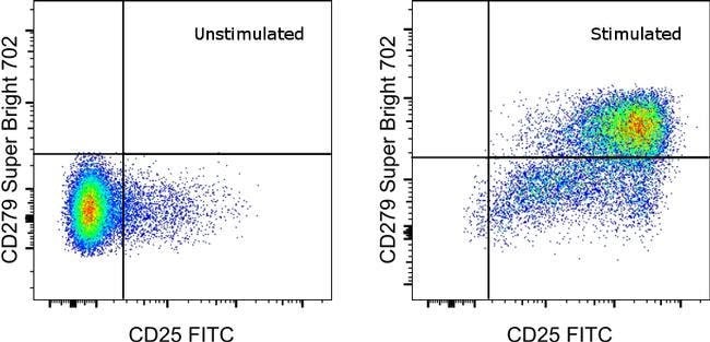
Description: The J105 monoclonal antibody reacts with the human PD-1 (programmed death-1), a 55 kDa member of the CD28 immunoglobulin superfamily. PD-1 contains the immunoreceptor tyrosine-based inhibitory motif (ITIM) and plays a key role in peripheral tolerance and autoimmune disease. PD-1 is expressed predominantly on activated T and B lymphocytes. Two novel members of the B7 family have been identified as the PD-1 ligands, PD-L1 (B7-H1) and PD-L2 (B7-DC). Evidence reported to date suggests overlapping functions for these two PD-1 ligands and their constitutive expression on some normal tissues and upregulation on activated antigen-presenting cells. Costaining experiments suggest that eBioJ105 recognizes a different epitope than MIH4 (Product # 11-9969-42). Applications Reported: This eBioJ105 (J105) antibody has been reported for use in flow cytometric analysis. Applications Tested: This eBioJ105 (J105) antibody has been pre-diluted and tested by flow cytometric analysis of stimulated normal human peripheral blood cells. This may be used at 5 μL (1.0 μg) per test. A test is defined as the amount (μg) of antibody that will stain a cell sample in a final volume of 100 μL. Cell number should be determined empirically but can range from 10^5 to 10^8 cells/test. Super Bright 702 is a tandem dye that can be excited with the violet laser line (405 nm) and emits at 702 nm. We recommend using a 710/50 bandpass filter. Please make sure that your instrument is capable...
Cell-mediated immune responses are initiated by T lymphocytes that are themselves stimulated by cognate peptides bound to MHC molecules on antig en-presenting cells (APC). T-cell activation is generally self-limited as activated T cells express receptors such as PD-1 (also known as PDCD-1) that mediate inhibitory signals from the APC. PD-1 can bind two different but related ligands, PDL-1 and PDL-2. Upon binding to either of these ligands, signals generated by PD-1 inhibit the activation of the immune response in the absence of 'danger signals' such as LPS or other molecules associated with bacteria or other pathogens. Evidence for this is seen in PD1-null mice who exhibit hyperactivated immune systems and autoimmune diseases. Despite its predicted molecular weight, PD-1 often migrates at higher molecular weight in SDS-PAGE.Specifications
| CD279 (PD-1) | |
| 5 μL/Test | |
| Flow Cytometry | |
| Super Bright 702 | |
| Mouse | |
| RUO | |
| PBS with BSA and 0.09% sodium azide; pH 7.2 | |
| Q15116 | |
| 100135775, 5133 | |
| Primary | |
| Affinity chromatography |
| eBioJ105 (J105) | |
| 4°C, store in dark, DO NOT FREEZE! | |
| Monoclonal | |
| Liquid | |
| IgG1 κ | |
| Human, Rhesus Macaque | |
| Pdcd1 | |
| CD279; EGK_05005; hPD1; hPD-1; hPD-l; hSLE1; Ly101; mPD-1; PD1; PD-1; Pdc1; Pdcd1; programmed cell death 1; programmed cell death 1 protein; programmed cell death protein 1; programmed cell death protein 1-like; programmed death 1; Protein PD1; protein PD-1; sCD279; SLEB2; soluble CD279; systemic lupus erythematosus susceptibility 2 | |
| Pdcd1 | |
| Antibody |
Spot an opportunity for improvement?Share a Content Correction
Product Content Correction
Your input is important to us. Please complete this form to provide feedback related to the content on this product.
Product Title