missing translation for 'onlineSavingsMsg'
Learn More
Learn More
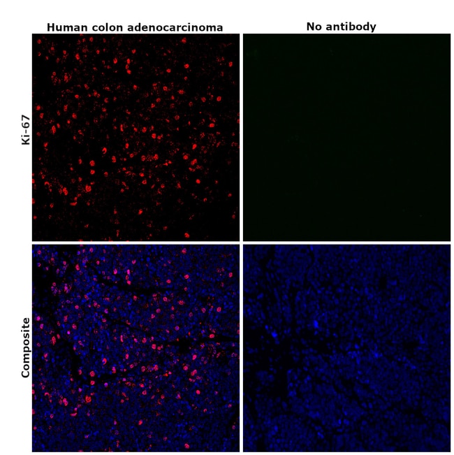
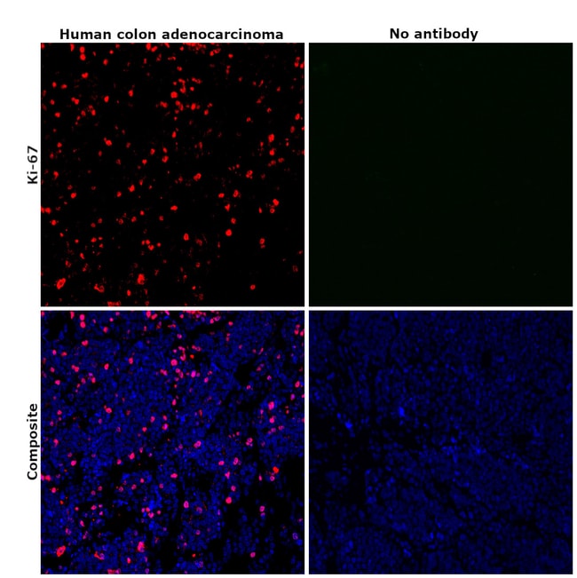
Invitrogen™ Ki-67 Monoclonal Antibody (20Raj1), eFluor™ 570, eBioscience™

Mouse Monoclonal Antibody
Brand: Invitrogen™ 41-5699-82
This item is not returnable.
View return policy
Description
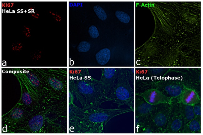
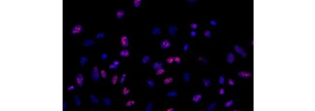
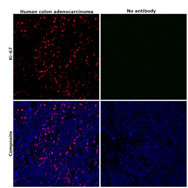
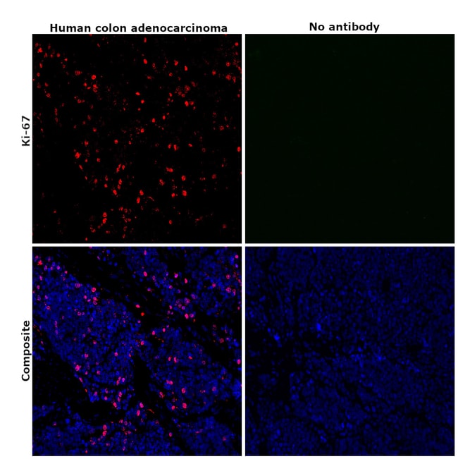
Description: The monoclonal antibody 20Raj1 recognizes the human Ki-67 protein. Two isoforms of Ki-67 exist, a 345 and 395 kDa form that are expressed in dividing cells. Ki-67 is expressed in all cell types and is detectable during active phases of the cell cycle (G1, S, G2, and mitosis) but is absent from resting cells (G0). During interphase, Ki-67 expression is localized to the nucleus but redistributes to the chromosomes during mitosis and has specifically been found to associate with heterochromatin-bound proteins such as chromobox protein homolog 3 (CBX3). In studies of tumor cells, Ki-67 expression has been used as a marker for determining the fraction of proliferating cells within a given population of tumor cells. This monoclonal antibody 20Raj1 recognizes canine Ki-67. Applications Reported: This 20Raj1 antibody has been reported for use in immunohistochemical staining of frozen tissue sections, immunohistochemical staining of formalin-fixed paraffin embedded tissue sections, and immunocytochemistry. Applications Tested: This 20Raj1 antibody has been tested by immunocytochemistry on fixed and permeabilized HeLa cells and can be used at less than or equal to 5 μg/mL. It is recommended that the antibody be carefully titrated for optimal performance in the assay of interest. Filter Recommendation: When using this eFluor™ 570 antibody conjugate, we recommend a filter that will capture the 570 emission wavelength (for example, Excitation 545/25, 565LP, Emi...
Ki-67 is a nuclear protein that is expressed during various stages in the cell cycle, particularly during late G1, S, G2, and M phases. The protein has a forkhead associated domain (FHA) through which it associates with euchromatin at the perichromosomal layer, the centromeric heterochromatin, and the nucleolus. Ki-67 is shown to have a cell cycle dependent topographical distribution with perinucleolar expression at G1, expression in the nuclear matrix at G2, and expression on the chromosomes during M phase. Ki-67 is commonly used as a proliferation marker because it is not detected in G0 cells, but increases steadily from G1 through mitosis. Ki-67 antibodies are useful in establishing the cell growing fraction in neoplasms. In neoplastic tissues, the prognostic value is comparable to the tritiated thymidine-labelling index. The correlation between low Ki-67 index and histologically low-grade tumors is strong. Ki-67 is routinely used as a neuronal marker of cell cycling and proliferation.Specifications
| Ki-67 | |
| Monoclonal | |
| 0.2 mg/mL | |
| PBS with 0.09% sodium azide; pH 7.2 | |
| P46013 | |
| Mki67 | |
| Affinity chromatography | |
| RUO | |
| 100686578, 4288 | |
| 4°C, store in dark, DO NOT FREEZE! | |
| Liquid |
| Immunohistochemistry (Frozen), Immunohistochemistry (Paraffin), Immunocytochemistry | |
| 20Raj1 | |
| eFluor 570 | |
| Mki67 | |
| antigen identified by monoclonal antibody Ki 67; antigen identified by monoclonal antibody Ki-67; Antigen identified by monoclonal antibody Ki-67 homolog; Antigen KI-67; Antigen KI-67 homolog; antigen KI-67; proliferation marker protein Ki-67; antigen KI-67-like; cb31; D630048A14Rik; I79_022666; Ki67; Ki-67; KIA; LOW QUALITY PROTEIN: proliferation marker protein Ki-67; marker of proliferation Ki-67; MIB-; MIB-1; Mki67; PPP1R105; Proliferation marker protein Ki-67; proliferation-related Ki-67 antigen; protein phosphatase 1, regulatory subunit 105; RP11-380J17.2; sb:cb31; si:ch211-250b22.7; unnamed protein product; wu:fa11g09; wu:fb57a07; wu:fi14e05 | |
| Mouse | |
| 100 μg | |
| Primary | |
| Canine, Human | |
| Antibody | |
| IgG1 κ |
Product Content Correction
Your input is important to us. Please complete this form to provide feedback related to the content on this product.
Product Title
Spot an opportunity for improvement?Share a Content Correction